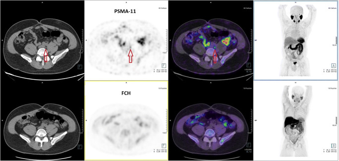
Fig. 3.
This 65-year-old patient underwent total prostatectomy. During follow-up, a rise in sPSA = 0.2 ng/mL prompted FCH PET/CT. a FCH PET/CT (lower row of images, from left to right: CT, PET, PET/CT fusion, MIP) was interpreted as negative for BCR. The FCH focus in the right lower part of the neck was interpreted as a probable hyperfunctioning parathyroid gland but no further exploration was performed. b PSMA-11 PET/CT (upper row of images) showed a significant 5-mm focus (arrow). Focused radiation therapy was performed on this lymph node and induced a drop in sPSA that persisted 1 year later, and the patient was accepted for a renal transplantation. This case illustrates the high sensitivity of PSMA-11 PET/CT to detect lymph node metastasis at sPSA as low as 0.2 ng/mL

