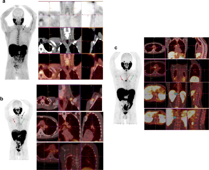
Fig. 4.
This patient with prostate cancer, Gleason 4 + 3, was treated by prostatectomy and radiotherapy. His first BCR after 2 years was treated by hormone therapy. His second BCR occurred 3 years later, sPSA = 1.2 ng/mL. a FCH PET/CT (from left to right: MIP, axial, coronal, and sagittal fused PET/CT slices) was then performed, showing a left focus of low intensity (SUVmax = 2.3) in a supraclavicular lymph node (black arrow), interpreted as equivocal. b On 68Ga-PSMA-11 PET/CT, a clear focus in the left supraclavicular lymph node (black arrow) was considered positive (SUVmax = 6.9), and 2 less intense foci were discovered in a left mediastinal lymph node (green arrow) (SUVmax = 2.6) and in the 7th right rib (red arrow) (SUVmax = 2.5), which were considered equivocal. Overall, the BCR was considered to be oligometastatic. Radiotherapy was performed on the left supraclavicular lymph node that induced a drop of sPSA. c Six months after the end of radiotherapy, sPSA increased at 4 ng/mL that prompted a follow-up PSMA-11 PET/CT. The irradiated left supraclavicular lymph node was no longer visible but the left mediastinal lymph node took up PSMA-11 more intensely (green arrow) and other mediastinal lymph nodes were now visible (SUVmax = 11.4) (brown arrow). The focus in the right rib was also more intense (SUVmax = 3.2) (red arrow), and two foci were discovered in the lumbar spine (bold arrows on coronal slices). The patient was then treated by hormone therapy. This case illustrates the high sensitivity of PSMA-11 to detect invasion of lymph nodes and the fact that any non-physiologic PSMA-11 focus should be taken into consideration, even with a moderate SUVmax

