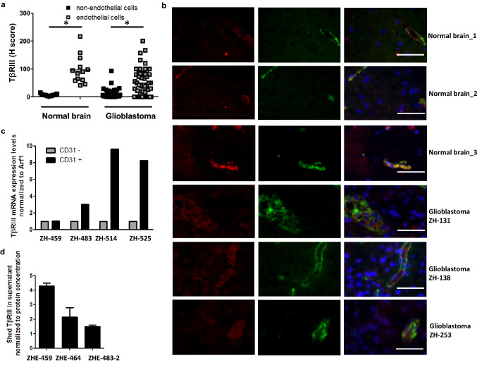
Fig. 1.
TβRIII levels in human glioblastoma in vivo. a TβRIII was detected immunohistochemically and quantified using the H-score in a TMA comprising 13 normal brain tissue samples obtained from epilepsy surgery and in tissue sections of 57 newly diagnosed and nine recurrent glioblastoma patients. Non-endothelial and endothelial cells, which were identified topographically aligning tumor vessels, were analyzed and scored separately; mean values are marked by the line, *indicates significance p < 0.001. b Representative photomicrographs of tissue sections from a analyzed for expression of TβRIII (red) and the endothelial marker vWF (green) by immunofluorescence. Scale bar: 50 µm. DNA was stained with 4′,6-diamidino-2-phenylindole (DAPI). c TβRIII expression was determined in CD31+ versus CD31− cell fractions of freshly dissociated glioblastoma tissues by RT-PCR. d Supernatants from CD31+ (endothelial) cells of three freshly dissected glioblastomas were assessed by ELISA for levels of shed TβRIII. Data were normalized to protein concentration of supernatants. (Color figure online)

