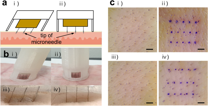
Figure 2.
Ex vivo skin penetration test using the MN devices. (a) Schematic illustration of the MN insertion into a porcine skin ex vivo by TMICS (i) and SMICS (ii). (b) Digital images of the MN penetration by TMICS (i) and SMICS (ii). TMICS MNs penetrated the skin with a tilted state (iii), and SMICS MNs penetrated vertically (iv). (c) Optical images of the skin before (i and iii), and after skin penetration by TMICS (ii) and SMICS (iv). The scale bars are 1 mm (n = 3).

