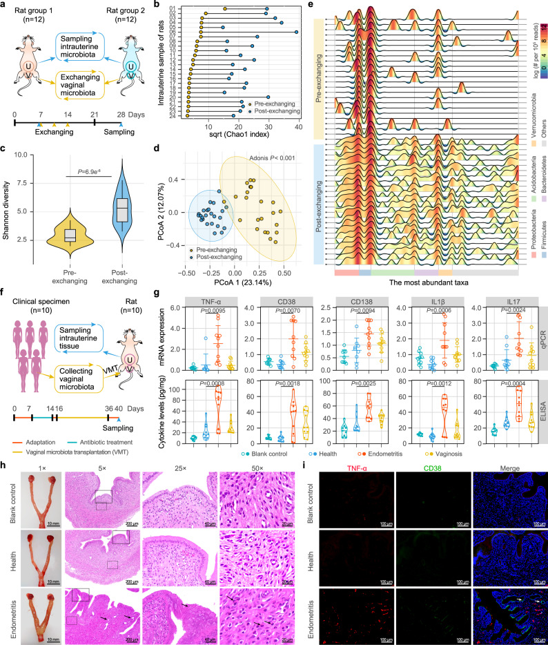
Fig. 4. Dysbiosis of the uterine microecology and inflammation of endometrial tissue triggered by vaginal perturbation.
a Study design of one-to-one exchange between the vaginal (V) microbiota of rats. Two groups of female Brown Norway (BN, n = 12) and Sprague Dawley (SD, n = 12) rats were used as donors and recipients, and the uterine (U) lavage fluid of each rat was sampled 3 weeks after exchange and was used for 16S rRNA gene sequencing. b Chao 1 index of the uterine microbiome of each rat before (Pre-) and after (Post-) exchanging the vaginal microbiota. c Shannon diversity index of the uterine microbiome in the pre- and post-exchange rats. The violin with box plot shows the median and interquartile range, and the width of the violin represents the density distribution. P values were determined by two-tailed Wilcoxon test. d Principal coordinate analysis (PCoA) of the uterine samples of the rats before and after vaginal microbiota exchange. The significance of separation of two clusters was measured with the Adonis test (P < 0.05). e Variations in the diversity and structure of the uterine microbiome in the rats before and after exchange. The taxonomic classification and profiling are shown at the phylum level, with each wave represents a family. Color bar represents the relative abundance of each family per 105 reads. f Study design of vaginal microbiota transplantation (VMT) from women to rats. Vaginal lavage fluids were collected from 10 women each in the healthy, chronic endometritis and bacterial vaginosis groups and were transplanted into the vagina of SD rats (n = 10) after 1 week of antibiotic treatment. g The mRNA expression and cytokine levels of inflammatory factors in the endometrial tissues of VMT rats. For each group, n = 10. The upper, center, and lower lines among points show the means ± s.e.m in mRNA expression. The width of the violin represents the density distribution of cytokine levels, and the upper, center and lower lines among points represent upper quartile, median and lower quartile, respectively. P values were determined by two-tailed Student’s t-test. h The edematous uterine body and symptoms of the endometrial tissues in the VMT rats. The 25× and 50× fields of view show the areas within the solid and dashed frames of the 5× field of view. The black arrows under 5×, 25×, and 50× fields of view show hyperplastic glands, endometrial epithelium, and inflammatory granulocytes in the uterus of rats transplanted with the vaginal microbiota of women with endometritis, respectively. i Immunofluorescence assay illustrating the TNF-α and CD38 signals in the endometrial tissue of the VMT rats. The white arrow shows signals from inflammatory factors. For h, i images are representative of three independent experiments with similar results.

