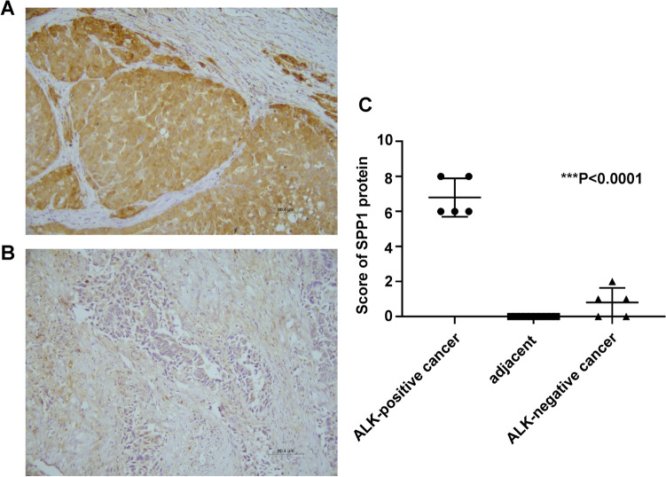
Figure 5.
Immunohistochemical staining of SPP1. (A) ALK-positive lung cancer. (B) ALK-negative lung cancer. Magnification, × 200, scale bars: 90.4 µm. (C) The scores were determined by evaluating the percentage and intensity of immunopositivity and were analyzed by paired-samples t test. There was a significant upregulation of SPP1 expression in ALK-positive lung cancers compared to ALK-negative lung cancers and paired adjacent normal tissues (**P < 0.0001).

