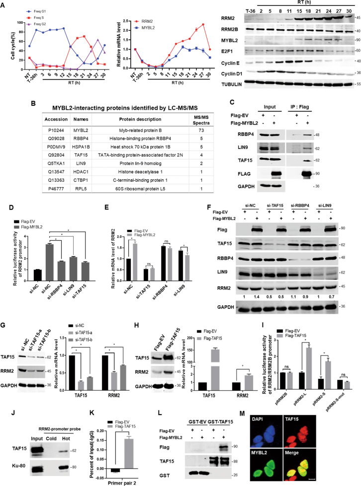
Fig. 4. MYBL2-related complex contributed to RRM2 transcription during S-phase in CRC cells.
A DLD1 cells were synchronized by 40uM lovastatin treatment for 36 h, and then released for analyses at the indicated time points. Left, FACS analyses of cell cycle phases (NT nontreatment, T treatment with lovastatin, RT remove treatment); Middle, qPCR for RRM2 and MYBL2; Right, western blotting for RRM2, RRM2B, MYBL2, E2F1, Cyclin E, Cyclin D1, and TUBULIN (as loading control). B DLD1 cells were transfected with empty vector (EV) or Flag-MYBL2 expression plasmid for 48 h, the cell lysates were co-immunoprecipitated with anti-Flag antibody, and MYBL2-interacting proteins were identified by LC-MS/MS. C The above MYBL2-interacting proteins were validated by western blotting with antibodies against RBBP4, LIN9, TAF15, FLAG, and GAPDH (as loading control). D The effects of knockdown of RBBP4, LIN9, or TAF15 on the activity of RRM2 promoter reporter (pRRM2-S) upregulated by MYBL2 transfection in DLD1 cells. E, F DLD1 cells were transfected with the siRNAs of negative control, TAF15, RBBP4, and LIN9, respectively, and then transfected with empty vector (EV) or MYBL2 expression plasmid for 48 h. qRT-PCR and western blotting were performed for analyses as indicated. G, H The effects of overexpression or knockdown of TAF15 on the mRNA and protein levels of RRM2 were analyzed by qRT-PCR (normalized by actin) and western blotting (GAPDH as loading control), respectively, in HCT116 cells. I Relative luciferase reporter activity of the truncated or mutated RRM2 promoter while co-transfected with TAF15 expression plasmid for 48 h in HCT116 cells, pRL-SV40 as an internal control reporter. J Nuclear proteins obtained from HCT116 cells were pulled down by a non-biotin-labeled (cold) or biotin-labeled (hot) DNA probe of RRM2 promoter (−2465/+23), western blotting was used to detect the biding of TAF15 and Ku80 (as binding control). K Chromatin extracted from HCT116 cells was immunoprecipitated with the Flag or IgG antibodies, qRT-PCR was carried out on immunoprecipitated DNAs using the primer pair2 as shown in Fig. 3A. L The whole lysates of HCT116 cells transfected with Flag-EV or Flag-MYBL2 were pulled down by GST-TAF15 or GST and then analyzed by immunoblotting. M HCT116 cells were plated into coverslip in six-well plate and then immunofluorescent staining was performed with antibody against TAF15 (red) or MYBL2 (green), scale bars: 10 μm.

