Figure 2.
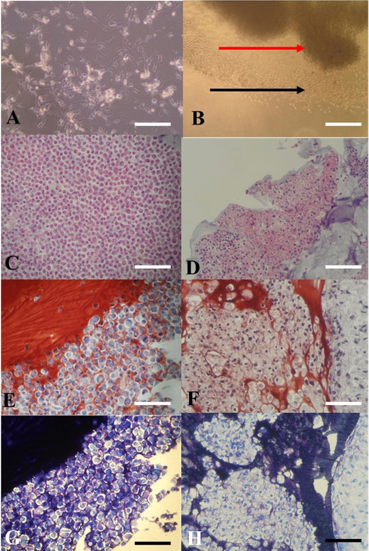
(A,B) In vitro culture images: (A) Chondrocytes in two-dimensional (2D) culture de-differentiating into fibroblast-like cells; (B) In vitro cultured chondrocytes growing in a tissue-like manner in three-dimensional (3D) thermo-reversible gelation polymer (3D-TGP) culture (the red arrow indicates the tissue; the black arrow indicates the cells migrating out into the 3D environment into the tissue); (C,D) H-and-E staining images: (C) Chondrocytes in 2D observed as individual cells. (D) 3D-TGP tissue-engineered chondrocytes exhibiting continuous tissue morphology with hyaline phenotype; (E,F) Safranin O/Fast Green staining images: (E) Chondrocytes in 2D observed as individual cells. (F) 3D-TGP tissue-engineered chondrocytes exhibiting continuous tissue morphology; (G,H) Toluidine blue images: (E) Chondrocytes in 2D observed as individual cells. (F) 3D-TGP tissue-engineered chondrocytes exhibiting continuous tissue morphology (All scale bars = 100 μm).

