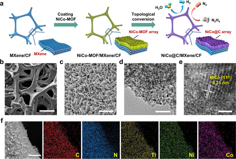
Fig. 2. Characterizations of NiCo@C/MXene/CF.
a Schematic illustration of the synthetic strategy of NiCo@C/MXene/CF. b SEM image showing macroporous scaffold of this electrode. Scale bar, 200 μm. c SEM image of mesoporous networks of NiCo@C nanosheets on electrode surface. Scale bar, 3 μm. d TEM image of a NiCo@C nanosheet. Scale bar, 100 nm. e HRTEM image of NiCo nanocrystallite on the nanosheet. Scale bar, 3 nm. f Elemental mapping showing the uniform distribution of C, N, Ti, Ni, and Co elements in this electrode. Scale bar, 5 μm.

