
Figure 1.
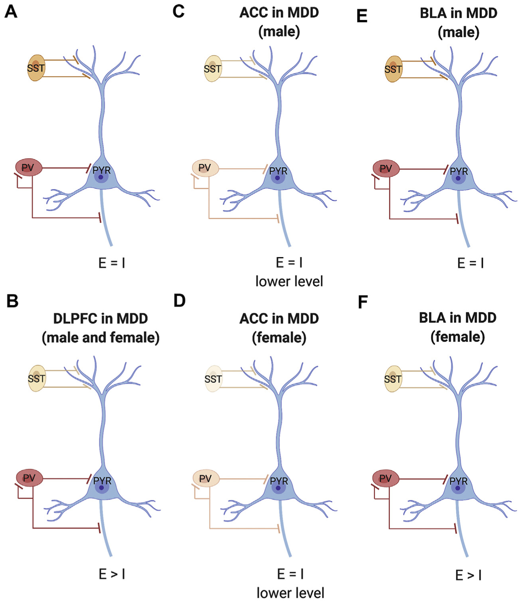
Schematic of neocortical microcircuitry and associated dysfunction in major depressive disorder (MDD). (A) Canonical microcircuitry in healthy control subjects with excitation (E)/inhibition (I) balance. (B) In the dorsolateral prefrontal cortex (DLPFC) of male and female subjects with MDD, there is lower expression of somatostatin (SST) (indicated by lighter color), but no change in parvalbumin (PV), suggesting reduced inhibition of input of pyramidal cells (PYR). (C) In the anterior cingulate cortex (ACC) of male subjects with MDD, there is moderately lower expression of both SST and PV, suggesting reduced inhibition of input and output of pyramidal cells, i.e., lower E/I balance. (D) In the ACC of female subjects with MDD, there is a robust reduction in expression of SST and moderately lower PV, suggesting strong inhibition of input and reduced inhibition of output of pyramidal cells, suggesting E/I imbalance. (E) In the basolateral amygdala (BLA) of male subjects with MDD, there are no differences in SST or PV, suggesting conserved E/I balance. (F) In the BLA of female subjects with MDD, there is reduced expression of SST, but not PV, suggesting reduced inhibition of input of pyramidal cells and altered E/I balance.