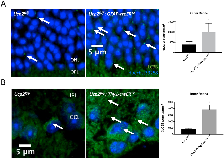
Figure 10. Layer Specific Ucp2-deletion dependent retinal autophagy.
We performed an immunohistochemistry experiment to label the autophagy marker LC3B (green) and DNA (Hoechst-33258, blue) in fixed frozen retinal tissue sections from Ucp2flox/flox (n=4), Ucp2flox/flox; Gfap-creERT2 (n=3), and Ucp2flox/flox; Thy1- creERT2 (n=3) mice. In mice expressing Gfap-creERT2, Ucp2 was deleted in GFAP-expressing astrocytes and müller glia, and in Thy1-creERT2-expressing mice, Ucp2 was deleted in projection neurons, which in the retina are retinal ganglion cells. Example images are on the left, and quantifications of LC3B puncta density are on the right. Compared to the same regions of Ucp2flox/flox retinas, LC3B puncta significantly increase in the outer retinas of Ucp2flox/flox; Gfap-creERT2, and in the inner retinas of Ucp2flox/flox; Thy1- creERT2 mice. *p<0.05.

