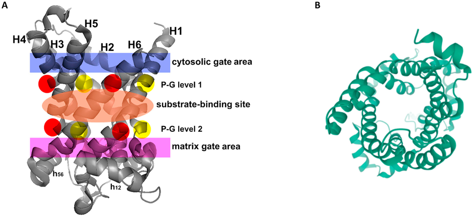
Figure 6.
(A) Crystal structure of the Adenine Nucleotide Transporter from which most of the structural models of UCPs are derived. The 3D crystal structure of the carboxyatractyloside-ADP/ATP carrier complex (devoid of the inhibitor) is shown with the following regions in color: cytosolic gate (blue); substrate-binding site (orange); and matrix gate (purple). In P-G level 1 and P-G level 2, prolines are shown in red and glycines in yellow. From (Palmieri & Pierri, 2010) with permission. (B) When the crystal structures are rotated 90 degrees and viewed end on, UCP2 and other members of the SLC25 transporter family have a clear central pore.

