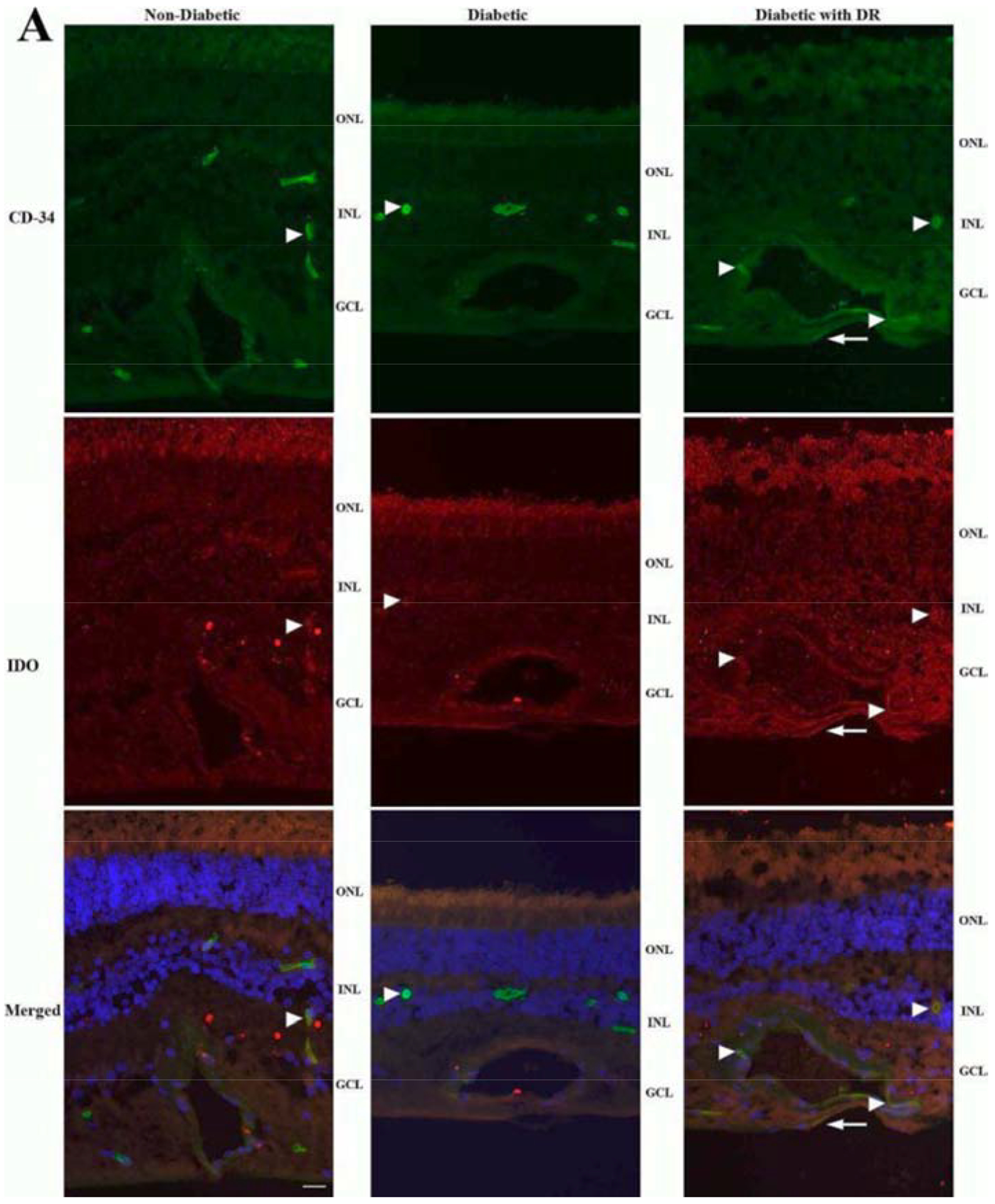
Figure 6. IDO expression is elevated in the retina of diabetic donors with DR.

Immunohistochemistry of retinal sections shows IDO (red) partially colocalizing with CD-34 (green) in the capillary endothelium (arrowheads) of the retina of diabetic donors with DR (right). IDO-positive staining was also observed in the capillary endothelium of the retina of diabetic donors without retinopathy (middle) and nondiabetic donors (left), but the signal appears diffuse and less intense (arrowheads). Additionally, some of the IDO signal in the inner retina of diabetic donors with retinopathy appears not to colocalize with CD-34 (arrows), suggesting expression in glial cells. Nuclei are stained with DAPI (blue). (Adapted from Nahomi et al., 2018)
