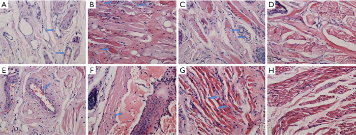
Figure 7.
Histopathological changes in the lips of rabbits on the 21st, 28th, and 35th days after injection of different mass concentrations of pingyangmycin in the experimental groups and after injection of normal saline in the control group by light microscope. (A, B, and C were respectively observed under light microscope on the 21st, 28th, and 35th days after injection of 8 mg/5 mL pingyangmycin; E, F and G were respectively observed under light microscope on the 21st, 28th, and 35th days after the injection of 8 mg/3 mL pingyangmycin; D and H were the manifestations under the light microscope on the 35th day after injection of normal saline in the control group; HE staining, magnification ×400). The arrows in (A) showed that the vascular endothelial cells were slightly edematous and the peripheral inflammatory cells were infiltrated. The arrows in (B) showed that hyaline degeneration of collagen fibers was observed and the thrombosis was found in some vascular lumens. The arrows in (C) showed that some capillary lumens were occluded by the thrombosis. The arrow in (E) showed that inflammatory cells infiltrated around vascular endothelial cells. The arrow in (F) showed that massive thrombosis and vascular lumens occlusion were observed in the blood vessels. The arrows in (G) showed that some muscle fibers were dissolved, broken and disordered. There was hyaline degeneration in the collagen fibers.

