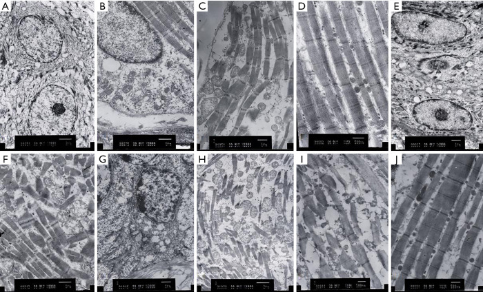
Figure 8.
Histopathological changes in the lips of rabbits on the 21st, 28th, and 35th days after injection of different mass concentrations of pingyangmycin in the experimental groups and after injection of normal saline in the control group by transmission electron microscope (TEM). (A and B, C, D were respectively observed under transmission electron microscope on the 21st, 28th, and 35th days after injection of 8 mg/5 mL pingyangmycin; F, G and H, and I were respectively observed under transmission electron microscope on the 21st, 28th, and 35th days after the injection of 8 mg/3 mL pingyangmycin; E and J were the manifestations under the transmission electron microscope on the 35th day after injection of normal saline in the control group; 80 kV, magnification ×1,000).

