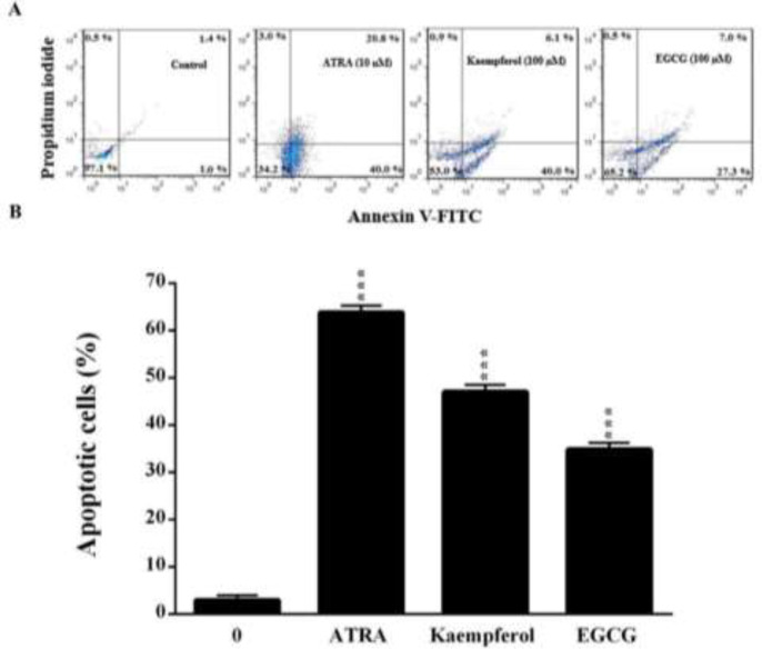
Figure 2.
The effects of kaempferol and EGCG on apoptosis in leukemia HL60 cells as evaluated by annexin V and propidium iodide double-staining. The cells were treated for five days with kaempferol and EGCG (100 µM) or ATRA. (A) A representative histogram of the fluorescence intensity of annexin V and PI double-stained cells (x-axis: green fluorescence of annexin-V-FITC indicating apoptotic cells; y-axis: red fluorescence of PI depicting necrotic cells). (B) Quantitative analysis of apoptosis as demonstrated in (A). The data are expressed as the mean±SEM of three independent experiments performed in triplicate. ***p<0.001 vs. the untreated control cells (concentration of 0).

