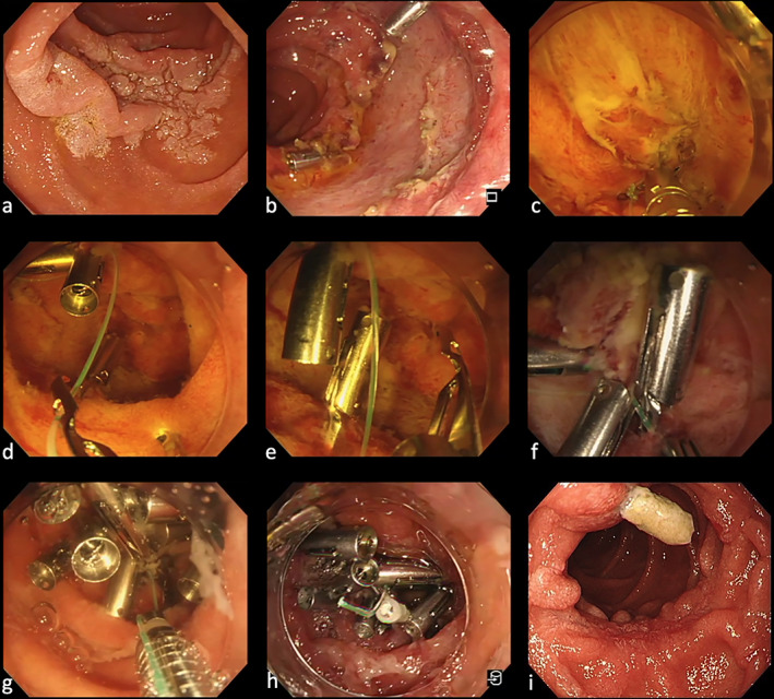
Figure 1.

(a) Paris, type 0‐IIa lesion of the descending portion of the duodenum. (b) Mucosal defect after duodenal ESD. (c) Place a reopenable‐clip fixed line on the oral mucosal defect. Next, a second clip grips line, the normal mucosa on the anal side of the mucosal defect margin and the muscular layer of the duodenal mucosal defect. (d) Using the reopenable‐clip over the line method (ROLM), a reopenable‐clip with a line passed through the tooth appears on the endoscope view. (e, f) By pulling the line slightly, the mucosal defect's margins are brought close to each other, and the mucosal defect's margins are fixed firmly. (g) Fixing the line with the modified locking‐clip technique before cutting to prevent it from slipping off the clip. (h) Completely closed mucosal defect. (i) The endoscopic image of the duodenal mucosal defect at the follow‐up after 2 months.
