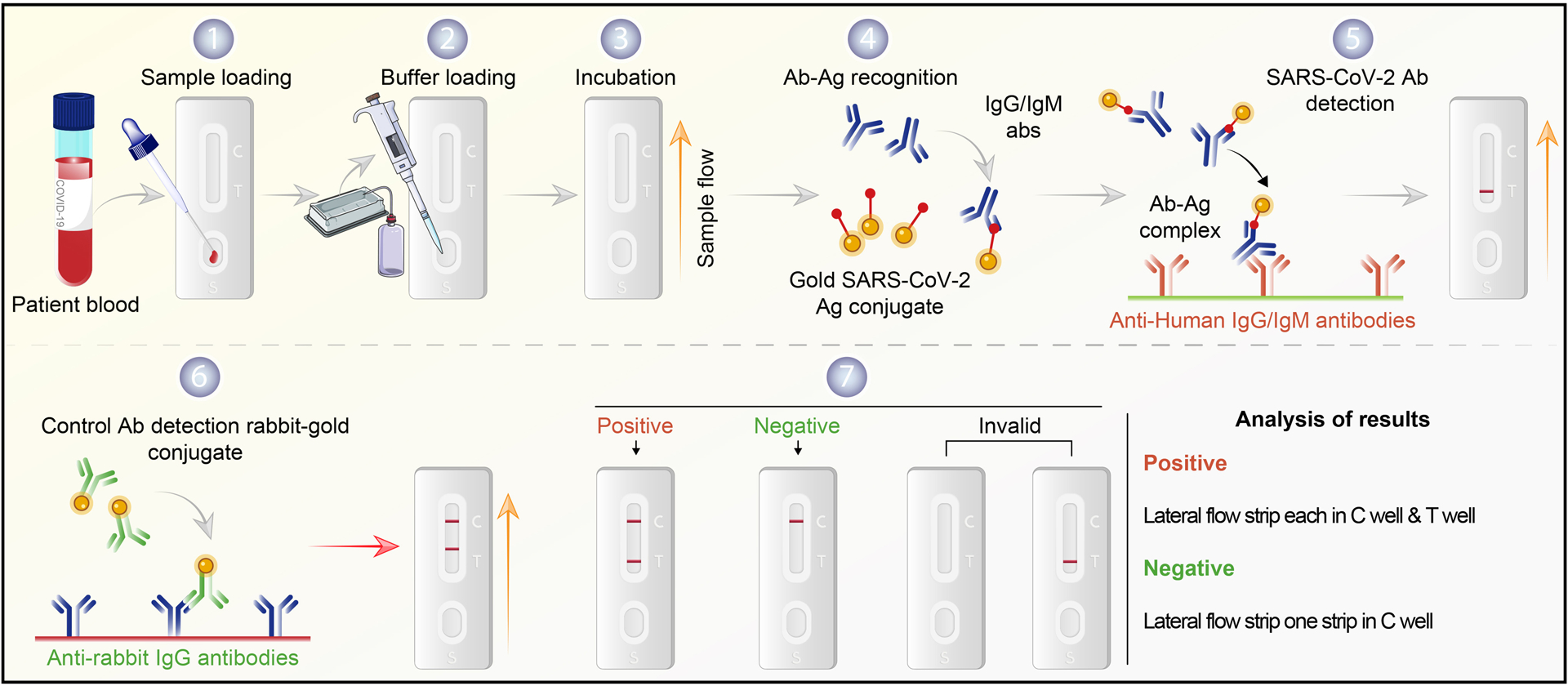
Figure 1. SARS-CoV-2 serological testing.

Commonly used immune-based tests contain SARS-CoV-2 specific recombinant antigens immobilized onto nitrocellulose membranes. Mouse anti-human IgM and IgG antibodies conjugated with colored latex beads are immobilized on conjugate pads. The test sample contacts the membrane within the test. The colored antibodies form latex conjugate complexes with human antiviral antibodies. This complex immobilized on the membrane is captured by the SARS-CoV-2 specific recombinant antigen. If SARS-CoV-2 virus-specific IgG/IgM are present in the sample, it leads to a colored band indicating a positive test result. The complex is captured on the membrane by goat anti-mouse antibody forming a red control line. A built-in control line appears in the test window. The absence of a colored band demonstrates a negative result. The workflow begins with patient serum added to the sample flow well. (1) Saline buffer is added dropwise (2) onto the serum sample (3) until the rabbit antibody-gold shows in the control (C) well. A positive test band indicates the presence of COVID-19 antibody (4–6). Results without a positive C band are invalid (7). Notably, this assay depicts a post-immune response and may show negative results for recently infected patients. It may also detect virus in previously infected but asymptomatic persons.
