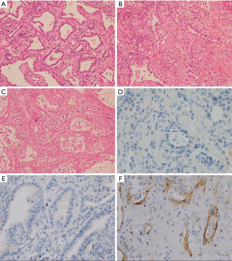
Figure 1.
Cellular changes with thermal ablation. Representative histologic findings (haematoxylin and eosin ×20) from Patient 3 demonstrating “viable” tumour (A) showed no effect from the ablation, with no evidence of necrosis or increase in inflammation. “Injured” tumour (B) showed increased inflammation in the form of inflammatory cells and/or fibrinous exudate, but no definite features of necrosis in the tumour cells. Injured tumour was typically seen at the interface of viable and necrotic tumour. “Necrotic” tumour (C) showed established necrosis with ghosted cell outlines, cytoplasmic eosinophilia, and nuclear hyperchromasia or dissolution. For Patient 5, PD-L1 tumour proportion score (TPS) was zero at baseline (D). Post ablation PD-L1 TPS was 5% in “viable” tumour (E) and 20% in “injured” tumour (F).

