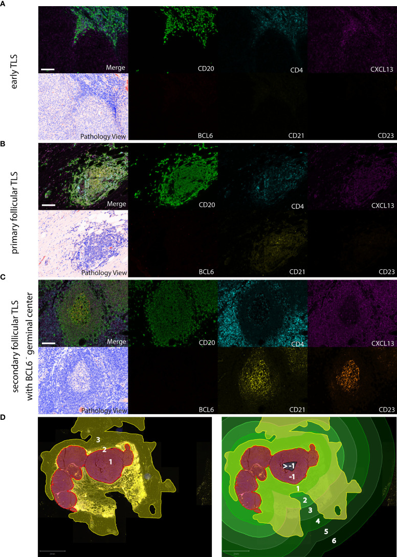
Figure 1.
Detection of different TLS phenotypes by 7-color multiplex immunohistochemistry in human melanoma tissues. Examples for (A) early TLS: dense CD20+ lymphocyte aggregates with CXCL13+ cells and the presence of some interspersed CD4+ T cells; (B) primary follicular TLS: CD20+ lymphocyte aggregates interspersed with CD4+ T cells and the presence of a CD21+ but CD23- dendritic network, surrounded by CXCL13 expressing cells; (C) secondary follicular TLS with a BCL6- germinal center: CD21+ and CD23+ dendritic networks within CD20+ lymphocyte aggregates with interspersed CD4+ T cells, surrounded by CXCL13 expressing cells. No accessory expression of BCL6 in lymphatic cells. Germinal centers are identifiable in routine light microscopy (see Pathology View, bottom left). An example for a BCL6+ secondary follicular TLS is given in Figure 5A . Images for each of the individual markers and their composites (for clarity without DAPI staining) are shown, together with the corresponding Pathology View (respective bottom left). Scale bars represent 100 µm. (D) Spatial annotation of tumor areas. Left: Definition of the invasive tumor front (red, 2) and the respective intra- and extratumoral compartments (brown, 1 and yellow, 3, respectively) in a tissue section of a human melanoma lymph node metastasis. Extratumoral compartments include site-specific and adipose tissue (yellow). Right: Allocation of intra- (brown/black) and extratumoral perimeters (green) of increasing radiuses (1 – max. 6 mm) drawn within and around the invasive tumor front. Counts and area of TLS were referred to the tissue area (yellow) within the respective intra- and extratumoral perimeters. TLS, tertiary lymphoid structures.

