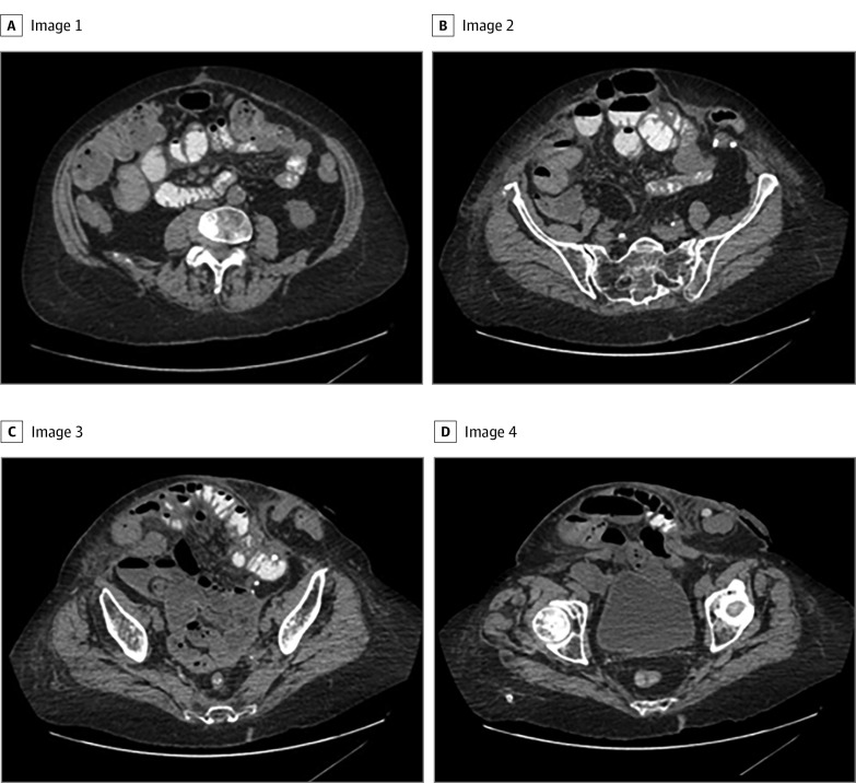
Figure 2. Representative Axial Computed Tomography Images of a Patient Requiring Component Separation.
To capture the entire hernia defect for this patient, a total of 22 images were included for development of the deep learning model. Panels A through D are 4 representative computed tomography cuts in order from cranial to caudal.

