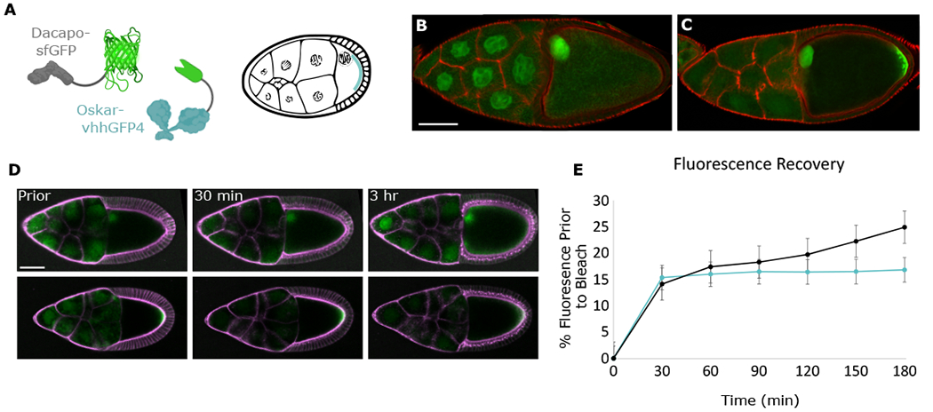
Figure 4. Oocyte-sourced Dap constitutes a major proportion of Dap in the NCs.

(A) Schematic of Osk-vhhGFP4 localized at the posterior of the egg chamber where it traps Dap-sfGFP.
(B and C) Confocal images of stage-10 Dap-sfGFP egg chambers without (B) or with (C) Osk-vhhGFP4. At this stage, Dap-sfGFP, like wild-type Dap (data not shown), has uniform levels in all NCs. Actin is stained with phalloidin (red).
(D) Time sequence of photobleach of NC region in Dap-sfGFP or Dap-sfGFP; Osk-vhhGFP4 egg chambers. Membranes were stained with CellMask Deep Red Plasma membrane Stain. Egg chambers were imaged prior to photobleaching of NC region and then imaged every 10 min for the next 3 h.
(E) Quantification of FRAP. Measurements of average sfGFP intensity of NC region, taken as percentage of average sfGFP intensity of NC region prior to bleach; n = 3 egg chambers for each condition. Error bars represent standard error. Scale bars, 70 μm (B) and (C), 50 μm (D).
