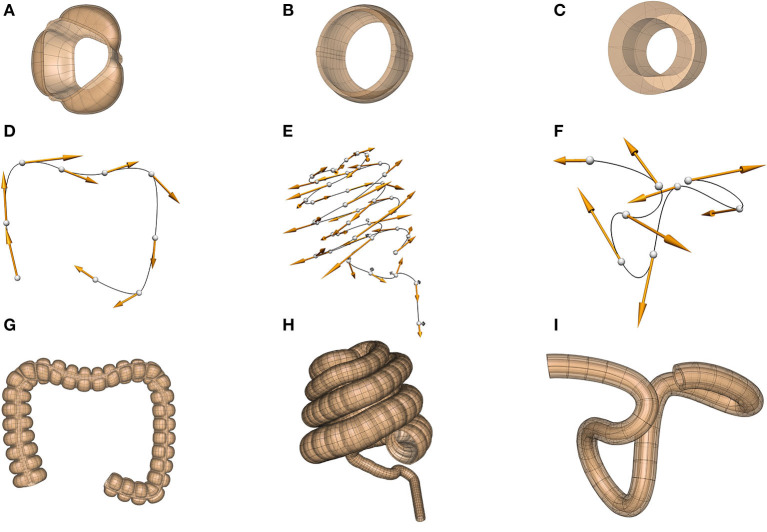
Figure 6.
Scaffolds for the colon in the human (dataset available from https://sparc.science/datasets/95), pig (dataset available from https://sparc.science/datasets/98), and mouse (dataset available from https://sparc.science/datasets/76). The scaffolds for each species are built in three steps: (A–C) define a sub-scaffold that captures the cross-sectional anatomy of the colon for the three species, (D–F) define the centerline of the colon, (G–I) attach these sub-scaffolds sequentially to the centerline to form the final scaffold.

