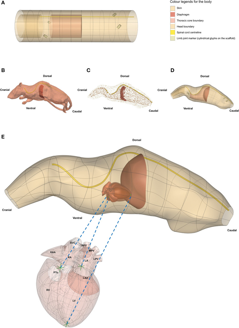
Figure 8.
Generating a whole rat body anatomical scaffold from data for integration with other organ scaffolds. (A) The 3D body coordinate system (dataset available from https://sparc.science/datasets/112) shows distinct anatomical regions with different colors. (B) The rat model (NeuroRat, IT'IS Foundation. 10.13099/VIP91106-04-0) in the vtk format with spinal cord and diaphragm visible. This model contains 179 segmented tissues with neuro-functionalized nerve trajectories (i.e., associated electrophysiological fiber models). (C) All tissue tissues from the rat body were resampled and converted into a data cloud to provide the means for generating the whole body scaffold. The data cloud is a 3D spatial representation of the tissue surface consisting of a certain density of points in the 3D Euclidean space. These points are used to define the objective function for the scaffold fitting procedure. The data cloud of the skin, inner core, spinal cord, and diaphragm are shown in (C) with the limbs and tail excluded. (D) The 3D body scaffold (A) was fitted to the rat data to generate the anatomical scaffold. (E) A generic rat heart scaffold was projected into the body scaffold using three corresponding fiducial landmarks (green arrows in heart and green spheres in the body). This transformation allows embedding of the organ scaffolds into their appropriate locations in the body scaffold, providing the required physical environment required for simulation and modeling. SCV, superior vena cava; RPV, right pulmonary vein; MPV, middle pulmonary vein; LPV, left pulmonary vein; RAA, right atrial appendage; RA, right atrium; LA, left atrium; LAA, left atrial appendage; PTo, pulmonary trunk outlet; RV, right ventricle; LV, left ventricle.

