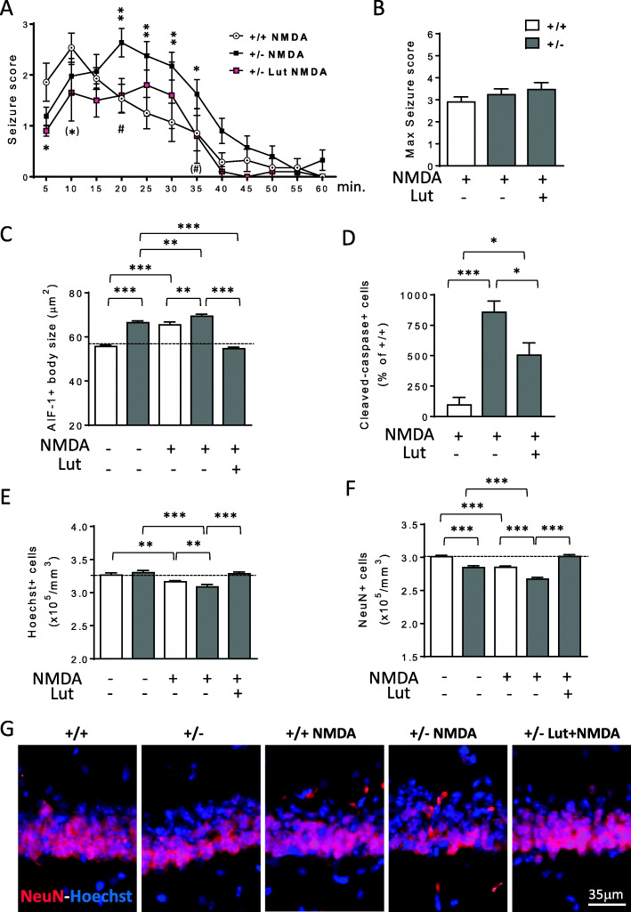
Fig. 10.
Effect of luteolin treatment on NMDA-induced excitotoxicity in the hippocampus of Cdkl5 +/- mice. A Graph represents seizure score of 3-month-old Cdkl5 +/+ (n=14) and Cdkl5 +/- (n=21) mice treated with a single intraperitoneal injection of NMDA (60 mg/kg) and of Cdkl5 +/- (n=10) mice treated with NMDA after 7 days of luteolin treatment. The results are presented as means ± SEM. (*) p=0.056; * p<0.05; ** p<0.01 as compared to the NMDA-treated Cdkl5 +/+ condition; (#) p=0.055; #p < 0.05; as compared to the NMDA-treated Cdkl5 +/- condition (Fisher’s LSD test after two-way ANOVA). B Histogram shows the mean of the maximum seizure score in mice treated as in A. C Mean AIF-1-cell body size of microglial cells in the hippocampus of Cdkl5 +/+ (n=5) and Cdkl5 +/- (n=6) mice treated with vehicle only, in Cdkl5 +/+ (n=3) and Cdkl5 +/- (n=3) mice treated with vehicle and NMDA and in Cdkl5 +/- (n=3) mice pre-treated for 7 days with luteolin before NMDA injection. Mice were sacrificed 8 days after NMDA treatment. D Number of cleaved caspase-3 positive cells in the hippocampus of Cdkl5 +/+ (n=3) and Cdkl5 +/- (n=4) mice treated with vehicle and NMDA, and in Cdkl5 +/- (n=6) mice pre-treated for 7 days with luteolin before NMDA injection. Mice were sacrificed 24 h after NMDA treatment. Data are given as a percentage of NMDA-treated Cdkl5 +/+ mice. E, F Quantification of Hoechst-positive cells (E), and NeuN positive cells (F) in CA1 layer of hippocampal sections from mice treated as in B. The results in B, E, and F are presented as means ± SEM. * p<0.05; ** p<0.01; ***p<0.001 (Fisher’s LSD test after one-way ANOVA). G Representative fluorescent images of hippocampal sections of mice treated as in B immunostained for NeuN and counterstained with Hoechst

