Fig. 3.
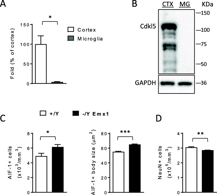
Non-cell autonomous microglial activation in the absence of Cdkl5. A Expression of Cdkl5 mRNA in cortex of 3-month-old Cdkl5 +/+ mice (n=3) and microglial cells purified from 3-month-old Cdkl5 +/+ mice (n=3). Data are given as a percentage of Cdkl5 cortical expression. B Example of immunoblot showing Cdkl5 and GAPDH levels in extracts from somatosensory cortex (CTX) of a wild-type Cdkl5 +/+ mouse and from microglial cells (MG) purified from Cdkl5 +/+ mice (n=4). C Quantification of AIF-1-positive cells (on the left) and mean cell body size (on the right) of microglial cells in hippocampal sections of wild-type (+/Y; n=4) and Emx1 KO (-/Y Emx1; n=5) mice. D Quantification of NeuN positive cells in CA1 layer of hippocampal sections of wild-type (+/Y; n=3) and Emx1 KO (-/Y Emx1; n=3) mice. The results in A, C, and D are presented as means ± SEM. * p<0.05; **p< 0.01; ***p<0.001 (two-tailed Student’s t-test)

