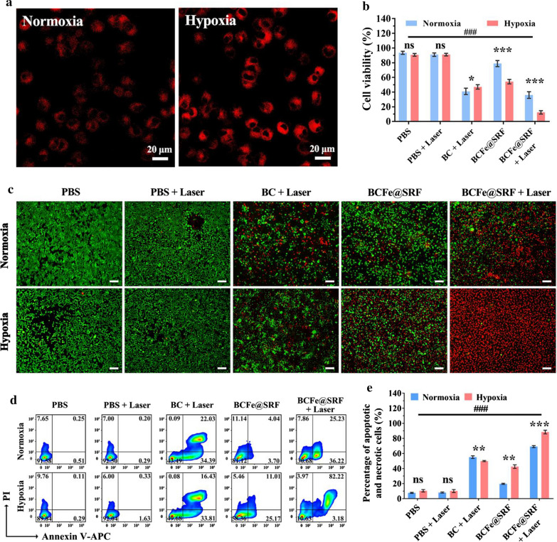
Fig. 4.
a CLSM images of hepa 1–6 cells treated with BCFe@SRF (Ce6 concentration: 1 μM) for 4 h in normoxic or hypoxia condition (Ce6: red, 405 nm laser excitation). b CCK-8 cell viability assay of hepa 1–6 cells treated with BCFe@SRF (Ce6 concentration: 1 μM) mediated PDT (670 nm light, 50 mW·cm−2, 5 min) in normoxic or hypoxia condition (*p < 0.05, **p < 0.01, ***p < 0.001, pairwise comparison; #p < 0.05, ##p < 0.01, ###p < 0.001, compared to the BCFe@SRF + Laser group in hypoxia condition, n = 5). c Live/dead staining assay (green: live cells; red: dead cells) and d annexin V-APC/PI apoptosis assay of hepa 1–6 cells treated with BCFe@SRF (Ce6 concentration: 1 μM) mediated PDT (670 nm light, 50 mW·cm−2, 5 min) in normoxic or hypoxia condition. e Percentage of apoptotic and necrotic cells in annexin V-APC/PI apoptosis assay (*p < 0.05, **p < 0.01, ***p < 0.001, pairwise comparison; #p < 0.05, ##p < 0.01, ###p < 0.001, compared to the BCFe@SRF + Laser group in hypoxia condition, n = 3)

