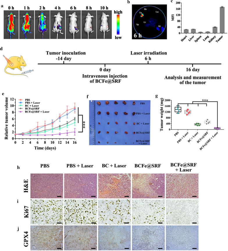
Fig. 6.
In vivo performance of BCFe@SRF in hepa 1–6 tumor bearing model (*p < 0.05, **p < 0.01, ***p < 0.001, n = 5). The involved laser irradiation (670 nm light, 0.4 W·cm−2, 5 min) was performed after 6 h of formulation injection. a In vivo fluorescence imaging of the mouse at different time points after intravenous injection of BCFe@SRF. b Ex vivo fluorescence imaging of the tumor and major organs harvested after 6 h of intravenous injection of BCFe@SRF. c The corresponding mean fluorescence intensity (MFI) of the harvested tumor and organs. d The schedule of the in vivo treatment for BCFe@SRF mediated synergistic therapy. e Time-dependent tumor growth curves. f The photographs and g average weights of the excited tumors at the end of the indicated treatment. h H&E, i Ki67 and j GPX4 immuno-histochemical staining of the dissected tumor after 24 h of the indicated treatment. Scale bar: 50 μm

