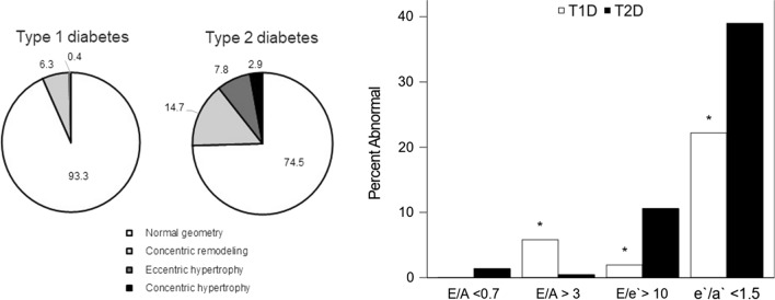
Fig. 1.
Left Ventricular Geometry and Diastolic Function by Diabetes Type. A. Distribution of Left Ventricular Geometry by Diabetes Type. Participants with type 1 and type 2 diabetes were stratified into four groups according to the LV mass cutoff > 51 g/m2.7 and relative wall thickness (RWT) cutoff of > 0.41: A Normal geometry (white), b concentric remodeling (increased RWT only, light gray), c eccentric hypertrophy (increased LVM only, dark gray), and d concentric hypertrophy (both increased LVM and RWT, black). p value difference between groups was < 0.0001. B Prevalence of Abnormal Diastolic Function by Diabetes Type. Diastolic function was compared to data from age similar healthy controls to assess the percent of abnormal diastolic function in participants with type 1 diabetes and type 2 diabetes [32, 33]

