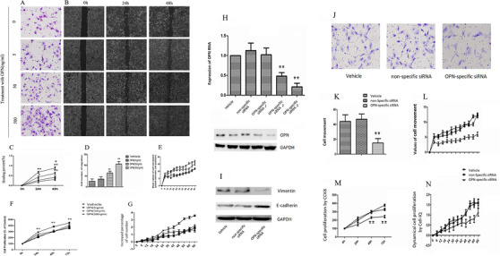
FIGURE 6.

Roles of OPN in lung cancer cell migration and proliferation. Cell number (A), migration (B), healing percentage (C), and number of migrated cells from upper to lower chambers in transwell (D) increased after OPN at 50 and 500 ng/ml. Dynamical cell proliferation was detected by Cell‐IQ monitoring (E) and CCK8 test (F), and dynamical cell movement by Cell‐IQ monitoring (G). Role of internal OPN in lung cancer cell proliferation was investigated by OPN‐specific siRNA, which inhibited the expression of OPN mRNA and production of OPN (H). Protein expression of high E‐cadherin and low vimentin were found in cellOPN− (I). We noticed cellOPN− had significantly lower capacity of cell movement from upper to lower chambers in transwell (J, K) and dynamical movement detected by Cell‐IQ monitoring (L), as well as cell proliferation by CCK8 (M) and by Cell‐IQ monitoring (N). Data are represented as mean ± SEM. Differences between groups were assessed by the Student's t‐test, after ANOVA analyses.* and ** stand for p‐values less than .05 and .01, in comparison with vehicle, and + and ++ stand for p‐values less than .05 and .01, as compared with OPN‐treated cells at 24 h, respectively
