Fig. 2.

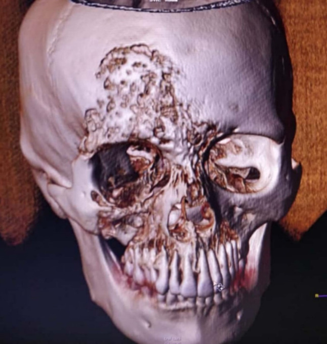
CT with 3D Reconstruction: Ill-defined lytic and sclerotic area with intermittent areas of bone destruction involving bilateral paranasal sinuses and maxilla, right frontal bone, orbit and zygoma

CT with 3D Reconstruction: Ill-defined lytic and sclerotic area with intermittent areas of bone destruction involving bilateral paranasal sinuses and maxilla, right frontal bone, orbit and zygoma