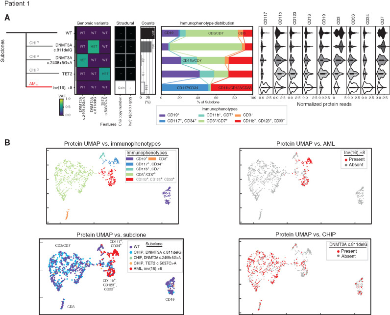
Figure 2.
Development of leukemia independent from clonal hematopoiesis. A, Clonal architecture as determined by scDNA and antibody–oligonucleotide sequencing in patient 1 shows the leukemic (AML, red line) clone developed separately from three clonal hematopoiesis (CHIP, gray lines) clones. Left: Genomic subclones with wild-type (WT), heterozygous (HET), present (Gain/+), or absent (-) features. Center: Immunophenotype as a percentage of each subclone. Right: Cell-surface protein expression for each subclone. B, For patient 1, cells clustered by cell-surface protein expression. UMAP shown colored by immunophenotype (top left) and genotype, with all subclones (bottom left), the leukemic subclone (top right), and one clonal hematopoiesis subclone (bottom right) depicted. VAF, variant allele frequency.

