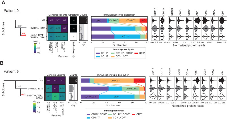
Figure 3.
Sequential development of leukemia from clonal hematopoiesis. Clonal architecture as determined by scDNA and antibody–oligonucleotidesequencing in patient 2 (A) and patient 3 (B) shows the leukemic (AML, red line) clone developed from clonal hematopoiesis (CHIP, gray line). Left: Genomic subclones with wild-type (WT), heterozygous (HET), present (+), or absent (–) features. Center: Immunophenotype as a percentage of each subclone. Right: Cell-surface protein expression for each subclone. VAF, variant allele frequency.

