Figure 4.

Spn binds to dying cells via PspA-GAPDH interaction
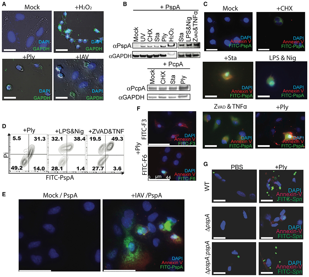
(A) A549 cells were incubated with H2O2, rPly, or influenza A virus (PR8, MOI 5). Surface-exposed GAPDH on unfixed cells was detected using mouse monoclonal anti-GAPDH antibody (green).
(B and C) Purified His6-PspA or PcpA proteins were incubated with healthy or dying cells (UV exposure; cycloheximide ; staurosporine; rPly; H2O2; lipopolysaccharide and nigericin; and zVAD-fmk, cycloheximide, and TNF-α). Bound PspA or PcpA were detected by (B) immunoblot and (C) fluorescent microscopy.
(D) Healthy or dying A549 cells were incubated with FITC-PspA. Adhered FITC-PspA to cells was analyzed by flow cytometry.
(E) A549 cells were infected with or without influenza A virus (PR8) and subsequently incubated with FITC-labeled PspA and Annexin V, then detected by fluorescent microscopy.
(F) Spn-infected A549 cells were incubated with F-3 or F-6, and Annexin V and PspA fragments bound to cells were detected by fluorescent microscopy.
(G) FITC-labeled Spn EF3030 (wild type [WT], pspA mutant [ΔpspA], or pspA revertant [ΔpspA:pspA]) were incubated with healthy or rPly-induced dying cells, and Spn adhesion was determined by fluorescent microscopy.
Data shown are representative of three independent experiments. Scale bars: 25 μm (A, C, and G); 44 μm (E and F).
