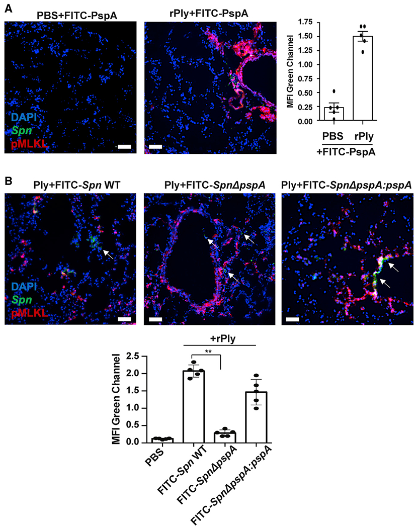
Figure 5. Spn binds to dying cell on mouse lung.

Mice were treated intratracheally with rPly (160 ng/mouse) or PBS. After 24 h, mice were challenged intratracheally with (A) FITC-rPspA or(B) 106 CFUs of FITC-labeled Spn EF3030 (WT, pspA mutant [ΔpspA], or pspA revertant [ΔpspA;pspA]) and then euthanized after 1 h (n = 5 per cohort). Lungs were collected and co-stained with anti-pMLKL (red) and DAPI (blue). White arrows indicate Spn. Mean fluorescence intensity (MFI) of the FITC-labeled rPspA (green) or Spn (green) was measured using ImageJ. Kruskal-Wallis test with Dunn’s multiple-comparison post-test. *p < 0.05, **p ≤ 0.01, ***p ≤ 0.001. Scale bars: 50 μm for all microscopic images.
