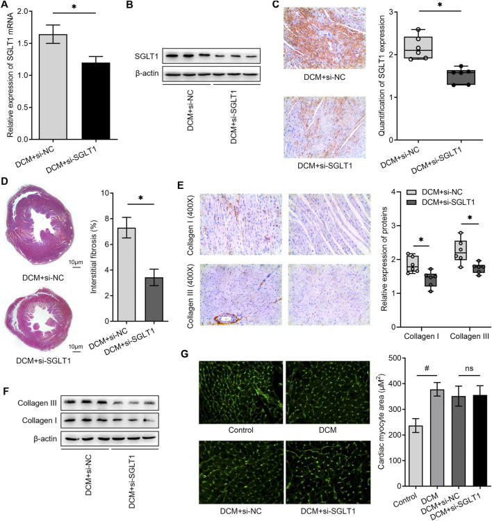
FIGURE 5.
SGLT1 inhibition alleviated myocardial fibrosis and apoptosis in diabetic rat hearts (n = 6). (A, B) Knockdown of SGLT1 was achieved by continuous intravenous injection into the tail vein with specific SGLT1 siRNA in DCM rats, and the knockdown efficiency was identified by detecting SGLT1 expression in myocardial tissues using RT-qPCR and western blotting analysis. (C) SGLT1 protein levels in myocardial tissue were measured by immunohistochemistry staining. (D) Masson’s trichrome staining of myocardial tissue shows interstitial fibrosis in DCM rats treated with si-NC or si-SGLT1. (E, F) Relative protein levels of collagen I and collagen III in myocardial tissue were measured using immunohistochemical staining and western blotting analysis (n = 6). (G) Wheat germ agglutinin staining and quantitation of myocardial cell size in DCM rats treated with si-NC or si-SGLT1 (n = 6).

