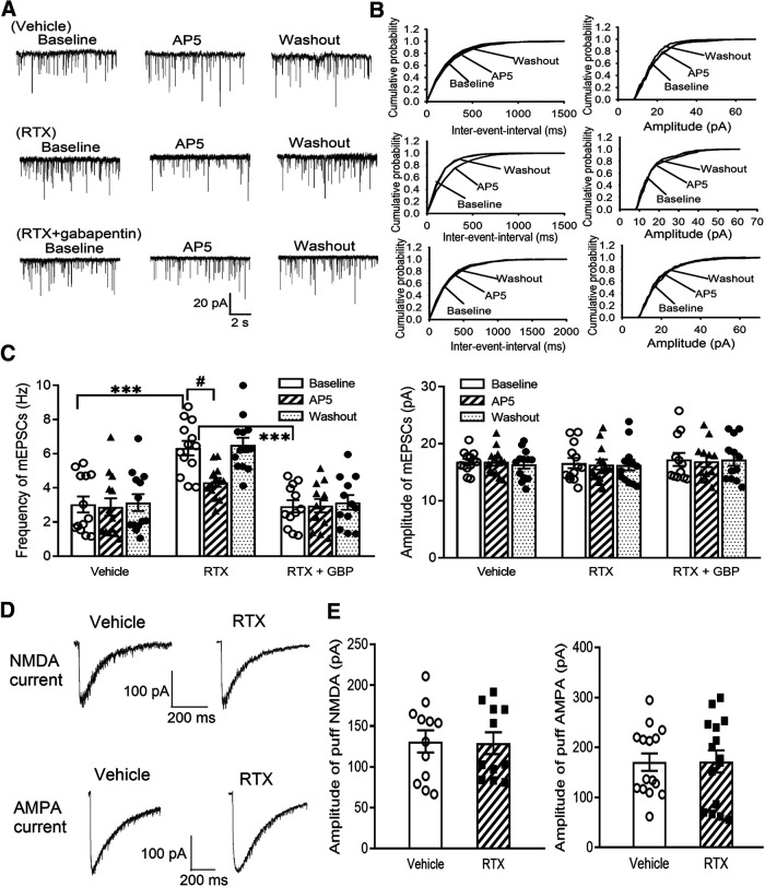
Figure 5.
RTX treatment increases glutamatergic input to spinal dorsal horn neurons via α2δ-1 and tonic activation of presynaptic NMDARs. A, B, Representative recording traces (A) and cumulative probability plots (B) show the effect of bath application of 50 μm AP-5 on mEPSCs of lamina II neurons from vehicle-treated rats, RTX-treated rats, and RTX-treated rats from which spinal cord slices were incubated with 100 μm gabapentin. C, Summary data show the effect of AP-5 on the frequency and amplitude of mEPSCs of lamina II neurons from vehicle-treated rats (n = 13 neurons), RTX-treated rats (n = 13 neurons), and RTX-treated rats from which spinal cord slices were incubated with GBP (n = 12 neurons). D, E, Representative current traces (D) and mean data (E) show currents elicited by puff application of 100 μm NMDA (n = 12 neurons in the vehicle group; n = 11 neurons in the RTX group) or 20 μm AMPA (n = 15 neurons per group) onto lamina II neurons from vehicle-treated rats and RTX-treated rats. Data are means ± SEM. ***p < 0.001 compared with baseline in RTX-treated rats. #p < 0.05 compared with baseline before AP-5 application. One-way ANOVA followed by Tukey's post hoc test.

