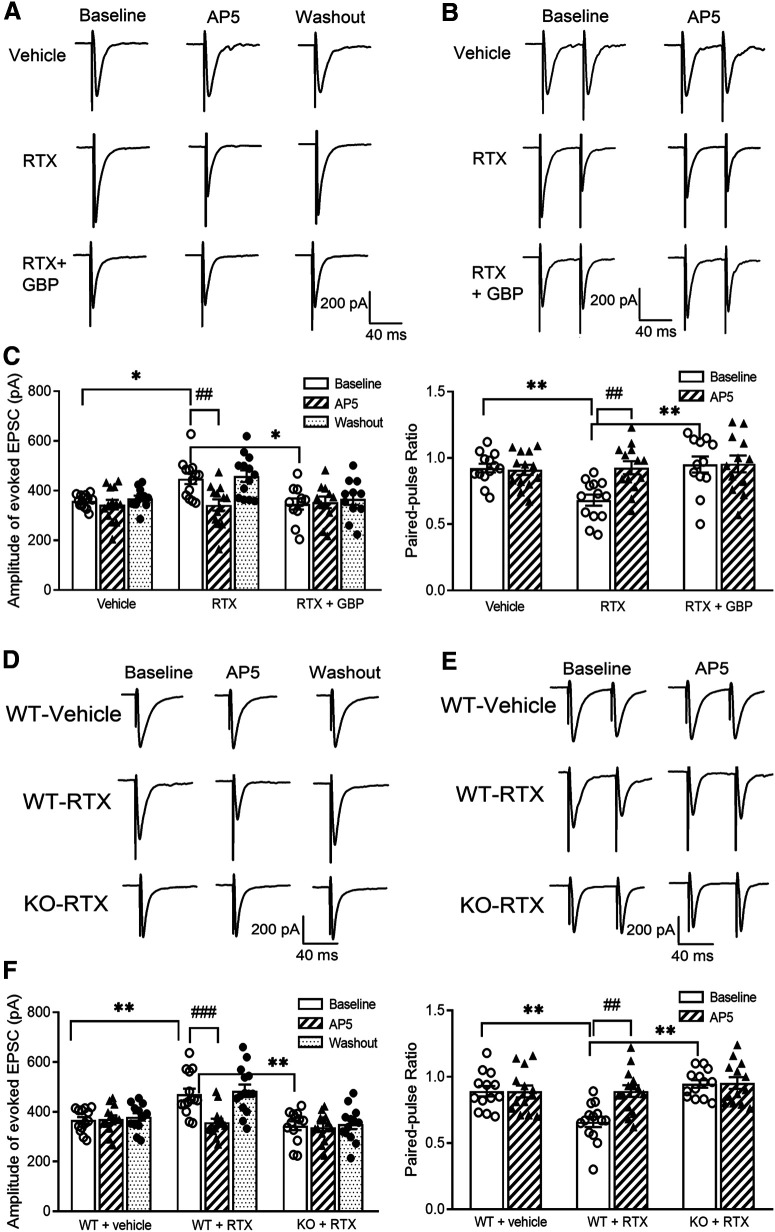
Figure 7.
RTX treatment augments glutamatergic input to spinal dorsal horn neurons via α2δ-1 and NMDARs present at primary afferent terminals. A, B, Original recording traces show the effect of 50 μm AP-5 on evoked monosynaptic EPSCs (A) and PPR (B) in lamina II neurons from vehicle-treated rats, RTX-treated rats, and RTX-treated rats from which spinal cord slices were incubated with 100 μm GBP. C, Summary data show the effect of AP-5 on the amplitude of evoked EPSCs and PPR in lamina II neurons from vehicle-treated rats (n = 13 neurons), RTX-treated rats (n = 13 neurons), and RTX-treated rats from which spinal cord slices were incubated with gabapentin (n = 12 neurons). D, E, Representative recording traces show the effect of 50 μm AP-5 on evoked monosynaptic EPSCs (D) and paired-pulse evoked EPSCs (E) in lamina II neurons from vehicle-treated WT mice, RTX-treated WT mice, and RTX-treated Cacna2d1-KO mice. F, Mean data show the effect of AP-5 on the amplitude of evoked EPSCs and paired-pulse ratio in lamina II neurons from vehicle-treated WT mice (n = 13 neurons), RTX-treated WT mice (n = 14 neurons), and RTX-treated Cacna2d1-KO mice (n = 13 neurons). Data are means ± SEM. *p < 0.05, **p < 0.01 compared with baseline in RTX-treated rats or WT mice. ##p < 0.01, ###p < 0.001 compared with baseline before AP-5 application. One-way ANOVA followed by Tukey's post hoc test.

