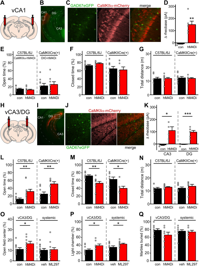
Figure 4.
Chemogenetic inhibition of excitatory neurons in the mouse vHPC. A, Schematic of bilateral targeting of vCA1. B, Representative image of the vHPC from a GAD67GFP(+) mouse receiving AAV8-CaMKIIα-mCherry in vCA1. Dashed lines highlight vCA1, vCA3, and vDG subregions. The white rectangle shows the area enlarged in panel C. C, Image of the vCA1 subregion showing GFP-positive (GABA) neurons within the area of viral infusion (left), CaMKIIα-driven mCherry expression (middle), and minimal overlap of GFP and mCherry fluorescence (right). D, Change in rheobase induced by CNO (10 μm) in control or hM4Di-expressing vCA1 pyramidal neurons (t(5) = 5.456, **p = 0.0025; unpaired Student's t test with Welch's correction; N = 2 mice and n = 6–8 total recordings/group). E, F, left, Percentage of time spent in open (t(17) = 0.8167, p = 0.4254; unpaired Student's t test) and closed (t(17) = 1.557, p = 0.1378; unpaired Student's t test) arms of the EPM by male C57BL/6J mice treated with intra-vCA1 hM4Di or control vector (N = 8–11 mice/group), following administration of CNO (2 mg/kg, i.p.). Right, Percentage of time spent in open (t(15) = 0.3685, p = 0.7177; unpaired Student's t test) and closed (t(15) = 0.3007, p = 0.7678; unpaired Student's t test) arms of the EPM by male CaMKIICre(+) mice treated with intra-vCA1 AAV8-hSyn-DIO-hM4Di(mCherry) or AAV8-hSyn-DIO-mCherry vector (N = 8–9 mice/group), following administration of CNO (2 mg/kg, i.p.). G, Total distance traveled during the EPM test by male C57BL/6J mice treated with hM4Di or control vector (left, t(17) = 0.09228, p = 0.9276; unpaired Student's t test), and in CaMKIICre(+) mice treated with intra-vCA1 DIO-hM4Di or control vector (right, t(15) = 0.3286, p = 0.7470; unpaired Student's t test). H, Schematic of bilateral targeting of vCA3/DG. I, Representative image of the vHPC from a GAD67GFP(+) mouse receiving AAV8-CaMKIIα-mCherry in the vCA3/DG subregion. The white open box shows the area enlarged in panel J. J, Image of the vCA3/DG subregion showing GFP-positive (GABA) neurons within the area of viral infusion (left), CaMKIIα-driven mCherry expression (middle), and minimal overlap of GFP and mCherry fluorescence (right). K, left, Change in rheobase induced by CNO (10 μm) in control or hM4Di-expressing vCA3 pyramidal neurons (t(3) = 3.749, *p = 0.0305; unpaired Student's t test with Welch's correction; N = 1–2 mice and n = 4–5 total recordings/group). Right, Change in rheobase induced by CNO (10 μm) in control or hM4Di-expressing vDG granule cells (t(6.5) = 7.298, ***p = 0.0002; unpaired Student's t test with Welch's correction; N = 2 mice and n = 7 total recordings/group). L, M, left, Percentage of time spent in open (t(14.84) = 3.074, **p = 0.0078; unpaired Student's t test with Welch's correction) and closed (t(17) = 3.228, **p = 0.0049; unpaired Student's t test) arms of the EPM by male C57BL/6J mice treated with intra-vCA3/DG hM4Di or control vector (N = 8–11 mice/group), following administration of CNO (2 mg/kg, i.p.). Right, Percentage of time spent in open (t(13) = 3.153, **p = 0.0076; unpaired Student's t test) and closed (t(13) = 2.795, *p = 0.0152; unpaired Student's t test) arms of the EPM by male CaMKIICre(+) mice treated with intra-vCA3/DG DIO-hM4Di or control vector (N = 7–8 mice/group), following administration of CNO (2 mg/kg, i.p.). N, Total distance traveled during the EPM test by male C57BL/6J mice treated with hM4Di or control vector (left, t(17) = 1.185, p = 0.2523; unpaired Student's t test) and in male CaMKIICre(+) mice treated with intra-vCA1 DIO-hM4Di or control vector (right, t(13) = 0.8588, p = 0.4060; unpaired Student's t test). O, left, Percentage of time spent in open-field center by male C57BL/6J mice treated with hM4Di or control vector (t(18) = 2.327, *p = 0.0318; unpaired Student's t test), following administration of CNO (2 mg/kg, i.p.). Right, Percentage of time spent in open-field center by male C57BL/6J mice treated with systemic ML297 (30 mg/kg, i.p.) or vehicle (t(22) = 0.1919, p = 0.8496; unpaired Student's t test) 30 min before testing. P, left, Percentage of time spent in light chamber of light-dark box by male C57BL/6J mice treated with hM4Di or control vector (t(18) = 2.767, *p = 0.0127; unpaired Student's t test), following administration of CNO (2 mg/kg, i.p.). Right, Percentage of time spent in light chamber by male C57BL/6J mice treated with systemic ML297 (30 mg/kg, i.p.) or vehicle (t(22) = 2.198, *p = 0.0387; unpaired Student's t test) 30 min before testing. Q, left, Percentage of marbles buried by male C57BL/6J mice treated with hM4Di or control vector (t(18) = 1.790, p = 0.0903; unpaired Student's t test), following administration of CNO (2 mg/kg, i.p.). Right, Percentage of marbles buried by male C57BL/6J mice treated with systemic ML297 (30 mg/kg, i.p.) or vehicle (t(22) = 0.4238, p = 0.6758; unpaired Student's t test) 30 min before testing.

