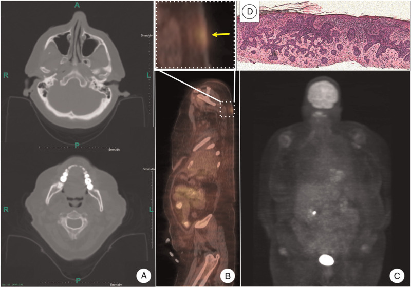
Figure 2.

Imaging studies and histopathology. A: Head and neck CT scan failed to show any skeletal or brain stem abnormalities suggestive of Gorlin syndrome. B: Whole body PET/CT showed no evidence of metastasis (lower panel). Large cutaneous tumor (yellow arrow) showed increased metabolic activity (upper panel). C: Brain and other internal organs showed physiologic distribution of fluorodeoxyglucose. D: Histomicrograph of the biopsy from the lesion shown in section proved to be a BCC (hematoxylin-eosin stain). BCC: Basal cell carcinoma; CT: Computed tomography; PET: Positron emission tomography.
