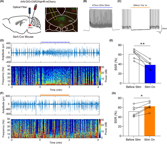
FIGURE 2.

Optical stimulation of DRN 5‐HT neurons affected the depth of isoflurane anesthesia. A, Left, schematic illustration of vius injection. Right, verification of virus expression and track of optical fiber (red, ChR2‐mCherry; green: Tph2). B,C, Confirmation of ChR2 or NpHR virus efficiency. D, Representative peri‐stimulation raw EEG and EEG spectra of blue laser. E, Activation of DRN 5‐HT neurons significantly reduced the BSR during anesthesia maintenance (61.26 ± 4.016% vs 38.26 ± 3.638%, P = 0.0015). F, Representative peri‐stimulation raw EEG and EEG spectra of yellow laser. G, Inhibition of DRN 5‐HT neurons significantly increased the BSR during anesthesia maintenance (51.98 ± 4.513% vs 62.53 ± 3.686%, P = 0.0191) (*P < 0.05, **P < 0.01, and ***P < 0.001, paired t test)
