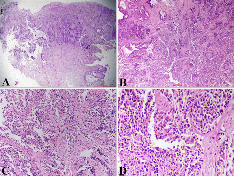
Figure 3. Hematoxylin and Eosin-Stained Sections From Cervical Tissue.
(A) Tumor with cervical epithelial ulceration (H&E, x20). (B, C) Stroma infiltrated by tumor cells arranged in diffuse sheets and vague nested pattern (H&E, x40, x100). (D) Monomorphic, small to round tumor cells having hyperchromatic nuclei, high nuclear-cytoplasmic ratio, and scant cytoplasm. Atypical mitosis is also noted (arrow) (H&E, x400).
H&E - Hematoxylin and eosin

