Figure 3.

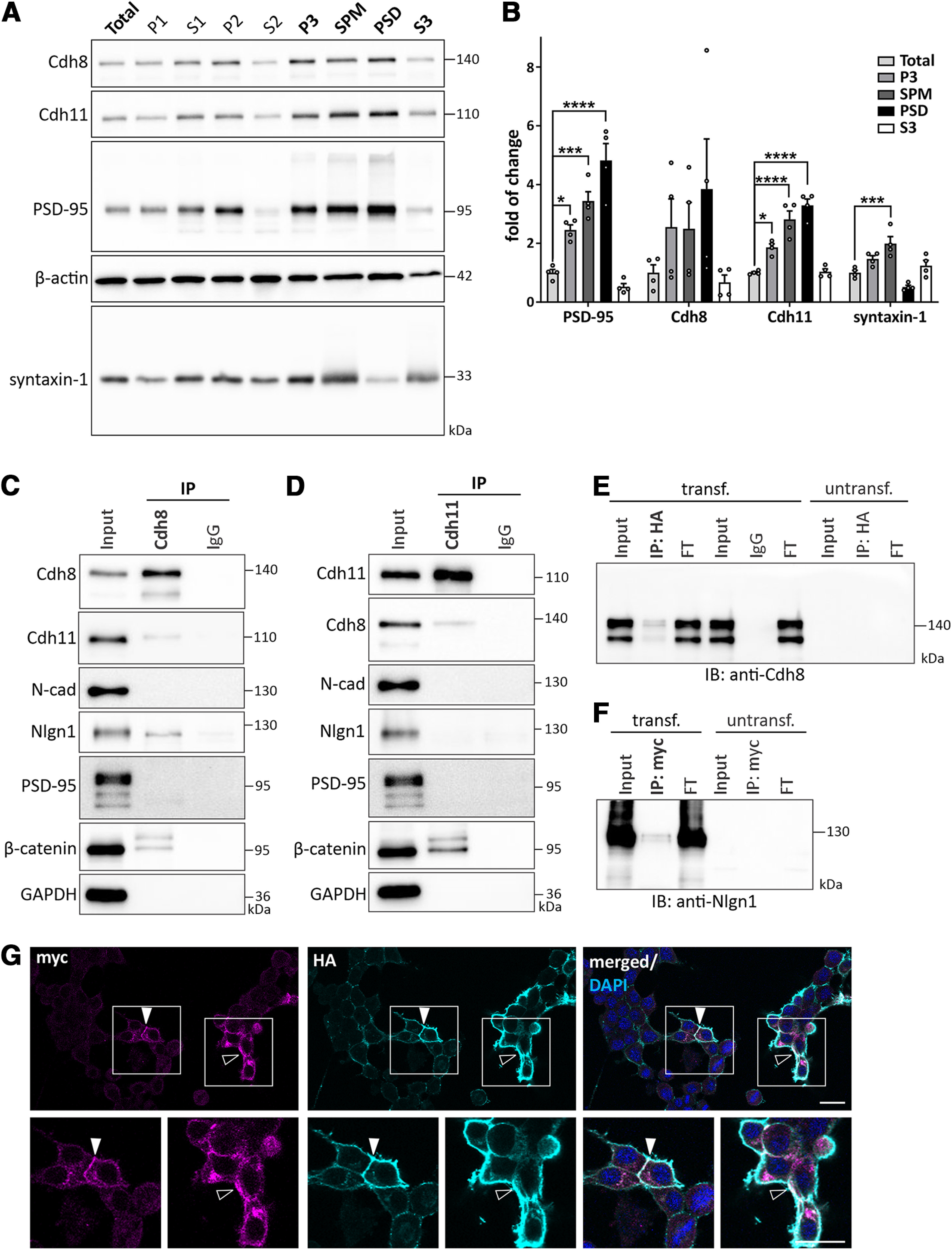
Cadherin-8 and cadherin-11 are expressed in synaptic compartments and cadherin-8 interacts with neuroligin-1. A, Forebrain tissues were subjected to synaptic fractionation analysis to determine the subcellular localization of cadherin-8 and cadherin-11. Markers (PSD-95 and syntaxin-1) were probed as control for purity of the fractionation. Total: total protein input, P1: nuclear, S1: cytosol/membranes, P2: crude synaptosome, S2: cytosol/light membranes, P3: synaptosome, SPM: synaptic plasma membrane, PSD: postsynaptic density, S3: synaptic vesicles. B, Quantification of protein enrichment in P3, SPM, PSD, and S3 fractions compared with total protein input. PSD-95: *p = 0.0159, ***p = 0.0002, ****p < 0.0001; Cdh8: p = 0.6618 (total vs SPM), p = 0.1673 (total vs PSD); Cdh11: *p = 0.0113, ****p < 0.0001; Syntaxin-1: ***p = 0.0009; one-way ANOVA with Dunnett’s multiple comparisons test. N = 8 P21 mice, two pooled forebrains per sample. Representative Western blotting of IP of (C) cadherin-8 and (D) cadherin-11 from P14 forebrain tissues. Cadherin-8 binds to cadherin-11, β-catenin, and neuroligin-1, whereas cadherin-11 binds to cadherin-8 and β-catenin, but not neuroligin-1. N = 3 independent co-IPs. Representative Western blotting of (E) IP of neuroligin-1-HA immunoblotted for Cdh8 and (F) IP of cadherin-8-myc immunoblotted for Nlgn1 from N2a cells overexpressing cadherin-8-myc and neuroligin-1-HA. Cadherin-8-myc is present in the IP of neuroligin-1-HA and neuroligin-1-HA is present in the IP of cadherin-8-myc. Cadherin-8 and neuroligin-1 are not expressed endogenously in N2a cells. FT, flow-through. N = 3 independent co-IPs. G, Immunocytochemistry of N2a cells overexpressing cadherin-8-myc (magenta) and neuroligin-1-HA (cyan), counterstained for DAPI (blue). Cadherin-8-myc and neuroligin-1-HA show partial co-localization at cell-cell contacts (arrowheads) and at the cell membrane (open arrowheads). Boxed areas are magnified. Scale bar: 20 μm.
