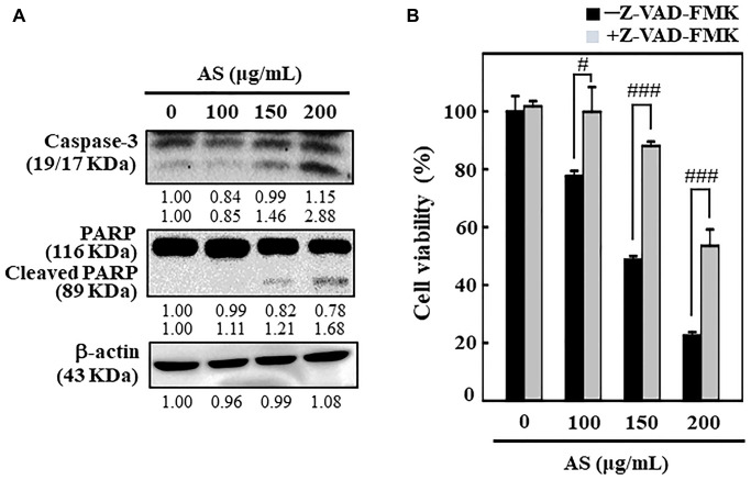
Figure 2.
AS induces apoptosis in colon cancer SW620 cells. The cells were treated with 100, 150, and 200 μg/mL of AS for 24 h. (A) Caspase-3 and PARP protein levels were inspected by Western blotting. Relative changes in protein bands were analyzed by commercially available quantitative software (AlphaEase, Genetic Technology Inc. Miami, FL, USA), with control as 1-fold. (B) Cells treatment was done with 20 μM Z-VAD-FMK, a caspase inhibitor, for 1 h then followed by 100, 150, and 200 μg/mL AS for 24 h. MTT assay was performed to assess cell viability. The data are expressed as the mean ± SD (n = 3) of three replicates. The statistical significance was defined as #p < 0.05 or ###p < 0.001 when compared with AS-treated cells.

