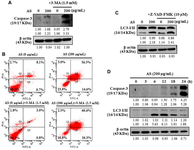
Figure 7.
Interplay between AS-induced autophagy and apoptosis in SW620 cells. (A) At first the cells were treated with or without 3-MA (1.5 mM) for 1 h, and incubated with AS (200 μg/mL) for 24 h. Caspase-3 cleavage was determined by Western blotting. (B) AnnexinV-FITC/PI staining was carried out to know about early/late apoptosis or necrosis. Flow cytometry analysis with or without 3-MA (1.5 mM) for 1 h, and incubated with AS (200 μg/mL) for 24 h. (C) The cells were pretreated with or without Z-VAD-FMK (10 μM) for 1 h, followed by incubation with AS (200 μg/mL) for 24 h. (D) Time-dependent AS (200 μg/mL for 0–24 h) effects of AS on Caspase-3 and LC3-I/II proteins were estimated by Western blotting. Relative changes in accord with the time were determined by commercially available quantitative software representing the control as 1-fold.

