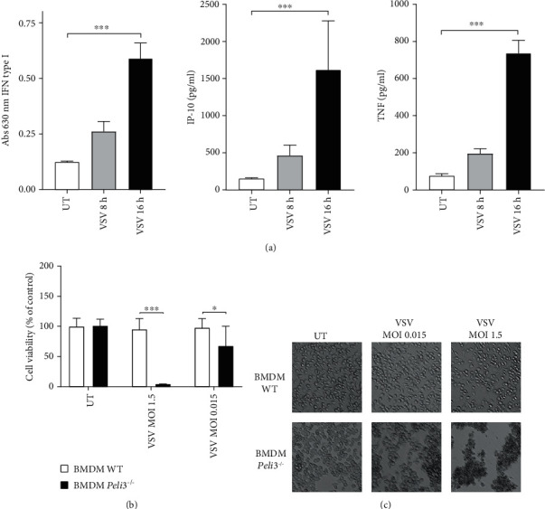
Figure 1.

Lack of Pellino3 results in VSV-induced disintegration of BMDMs. (a) WT BMDMs were infected with VSV at MOI 1.5. Cell supernatants were collected 8 and 16 h of postinfection. Type I IFN was measured by bioassay; level of IP-10 and TNF was measured by ELISA as described under Materials and Methods. (b, c) WT and Peli3−/− BMDMs were infected with VSV at MOI 1.5 and 0.015. Cell viability was evaluated by alamarBlue test 48 h of postinfection. Photographs were taken using 10x microscope objective. Results are representative of at least three independent experiments performed in triplicate (mean ± S.E.). Statistical analysis was carried out using the unpaired Student t-test using GraphPad Prism 7.04. p values of less than or equal to 0.05 were considered to indicate a statistically significant difference (∗p ≤ 0.05, ∗∗p ≤ 0.01, and ∗∗∗p ≤ 0.001).
