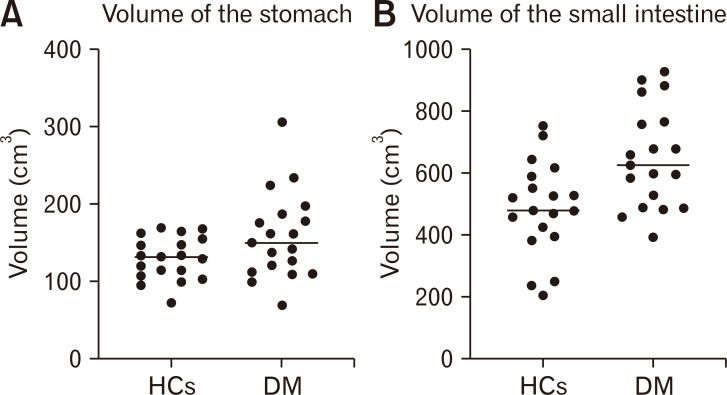
Figure 2.
Volumes of the gastric (A) and small intestinal walls (B) assesses with CT scans. The horizontal line shows the median. Diabetes patients had a significantly larger volume of the small intestinal wall (P = 0.003) but not of the stomach (P = 0.121). HCs, healthy controls; DM, patients with diabetes mellitus.

