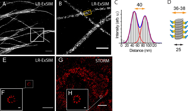
Figure S4.
LR-ExSIM of microtubules.(A) LR-ExSIM image of microtubules in a U2OS cell stained with antibody conjugated with NHS-MA-DIG anchors. (B) Magnification of A. (C) The transverse profile of the microtubule in the gold box in B. (D) Schematic of the structure of an immunostained microtubule. By fitting the peaks to Gaussian functions, we calculated the resolution (FWHM) of LR-ExSIM to be 34 nm. (E) LR-ExSIM image of Cep164 in distal appendages of a primary cilium of an expanded mouse embryonic fibroblast indirectly immunostained with NHS-MA-biotin secondary antibodies. The length expansion ratio is 4.2. (F) Magnified view of E. (G) STORM image of Cep164 in distal appendages of motile cilia of an unexpanded multiciliated mouse tracheal epithelial cell. (H) Magnified view of G. LR-ExSIM (E and F) and STORM (G and H) reveal structure of distal appendages with similar super resolution. The same primary antibody was used for both images. Scale bars, 1 µm (A), 500 nm (B), 2 µm (E and G), and 100 nm (F and H).

