Figure 2.

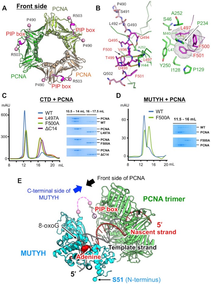
Structure and interactions of the complex between MUTYH and PCNA. (A) Overall structure of the PCNA trimer (green, lightgreen and lightorange) in complex with the PIP box (magenta) of MUTYH. (B) Interactions between the PIP box and PCNA. (C) Size-exclusion chromatography experiments using CTDs in complex with PCNA. Chromatograms of the wild type CTD (blue), L497A (red), F500A (green) and ΔC14 (purple), and SDS-PAGE analyses. WT indicates wild type. (D) Size-exclusion chromatography experiments using MUTYH (45–515) (wild type and F500A) in complex with PCNA. Chromatograms of the wild type (blue) and F500A (green), and SDS-PAGE analyses. (E) Structural model of replication-coupled repair by MUTYH (cyan and pink) and PCNA (green). MUTYH is loaded to DNA in the appropriate direction for the recognition and removal of adenine on the nascent DNA strand (red). The spacer region between the CTD and the PIP box (19 amino acids) is shown as a pink dashed line.
