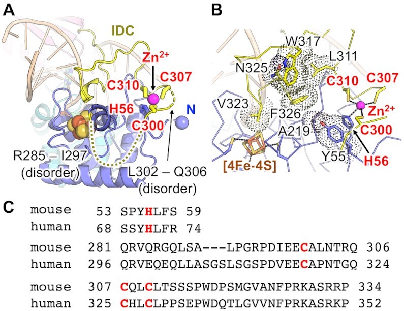
Figure 3.

IDC and Zn-binding motif. (A) Structure of the IDC and the Zn-binding motif. The amino acid residues Arg285–Ile297 and Leu302–Gln306 are disordered. (B) Van der Waals contacts observed in the IDC and [4Fe–4S] domain. (C) Amino acid sequences around the Zn-binding motif.
